√画像をダウンロード e coli under microscope 40x 498332-Can you see e coli under a microscope
Instead, their genetic material floats uncoveredIntroduction (E Coli) Commonly referred to as E coli, Escherichia coli is a bacterium that is typically found in a number of environments including various foods, soil and animal intestines E coli is very diverse and belongs to the genus Escherichia While most of the strains are harmless (and also important in the human intestinal tract), others are harmful and can cause very serious health implicationsE Coli Under The Microscope Types Techniques Gram Stain Escherichia Coli E Coli Microscope Bacterial Shape Coccus 1000x General Biology Lab Loyola Microscopy For The Winery Viticulture And Enology Onion Cell Under Microscope 4x 10x 40x
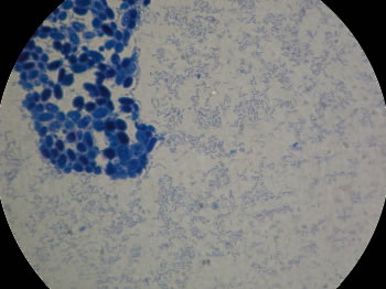
The Virtual Edge
Can you see e coli under a microscope
Can you see e coli under a microscope-Media in category "Microscopic images of Escherichia coli" The following 44 files are in this category, out of 44 total E coli 40Xjpg 2,592 × 1,944;4 Can a light microscope see bacteria?


What Does An E Coli Bacteria Look Like Under A Microscope Quora
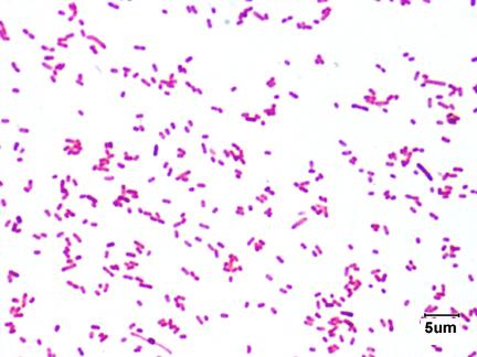
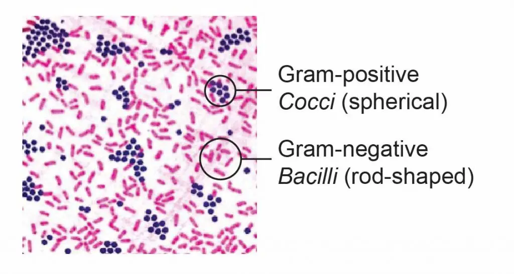
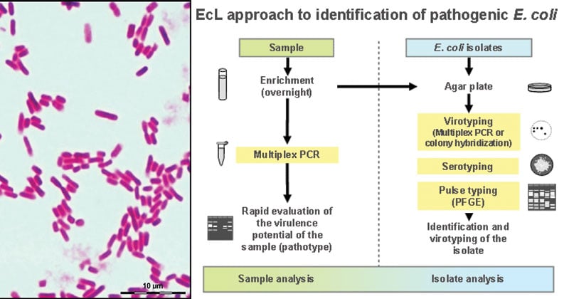
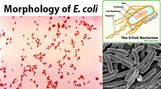
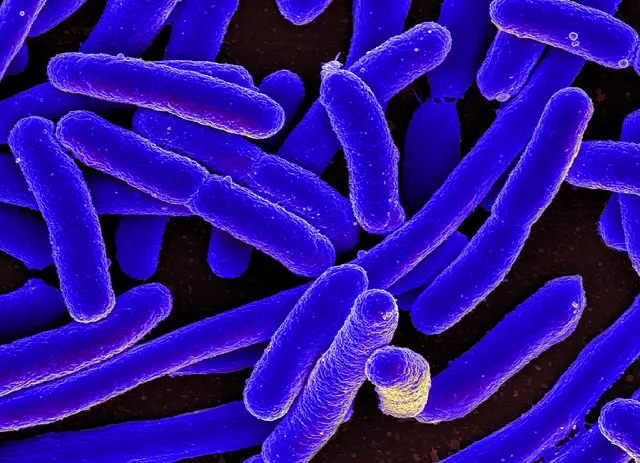
E coli is a Gramnegative rodshaped bacteria When Gram stained, the organism looks pink or red Here are a couple of pictures of a Gram stain of E coli that I did under the 100X objective lens on a standard light microscope You can of courseExamples of Bacteria Under the Microscope Escherichia coli Escherichia coli (Ecoli) is a common gramnegative bacterial species that is often one of the first ones to be observed by students Most strains of Ecoli are harmless to humans, but some are pathogens and are responsible for gastrointestinal infections They are a bacillus shaped bacteria that has a very fast growth (they can double every minutes), which is one of the main reasons they are used in research6 KB E coli Bacteria ()jpg 2,095 × 1,515;
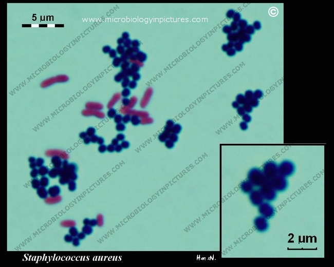
Gramstain Grampositive Microscopic appearance Cocci in grapelike clusters, diplococci, cocci Clinical significance Staphylococcus epidermidis is part of human skin flora (commensal) It can also be found in the mucous membranes and in animalsFor counting E coli using magnetic nanoparticle (MNP) probe under a darkfield in 30 min Results The antibodies functionalized MNP, binding to E coli to form a golden ringlike structure under a darkfield microscope, allowing for counting E coli This method via counting MNPconjugated E coli under darkfield microscopeMicroscopy and image processing Microscopy setup An inverted Eclipse TE00U (Nikon) microscope equipped with a DSQi1Mc (Nikon) (Table 1) monochrome digital cooled camera was used to collect brightfield or fluorescent images of culture samples through an SFluor 40x 09 NA oil/water (Nikon) objectiveThe proprietary Nis Elements Documentation v 4 software (Nikon) was used for image
Entamoeba coli is a nonpathogenic species of Entamoeba that frequently exists as a commensal parasite in the human gastrointestinal tract E coli (not to be confused with the bacterium Escherichia coli) is important in medicine because it can be confused during microscopic examination of stained stool specimens with the pathogenic Entamoeba histolytica1 Place slide on stage and secure with stage clip 2 Turn on the light and center the specimen over the light source 3 Focus specimen on low power using coarse focus adjustment 4 Use fine focus to sharpen the image under low power 5Yes, most of the bacteria range from 022 µm in diameter The length can range from 110 µm for filamentous or rodshaped bacteria The most wellknown bacteria E coli, their average size is ~15 µm in diameter and 26 µm in length As we talked above, you can see some bacteria in my cheek cells


Biol 230 Lab Manual Lab 1


Www Mccc Edu Hilkerd Documents Bio1lab3 Exp 4 Pdf
Microscopy and image processing Microscopy setup An inverted Eclipse TE00U (Nikon) microscope equipped with a DSQi1Mc (Nikon) (Table 1) monochrome digital cooled camera was used to collect brightfield or fluorescent images of culture samples through an SFluor 40x 09 NA oil/water (Nikon) objectiveThe proprietary Nis Elements Documentation v 4 software (Nikon) was used for imageE Coli Under Microscope Gram Stain Gram Negative Bacilli Or Gram Stain Gram Negative Pink Colored Small Rod Shape E Coli Under Light Bacterial Staining Onion Cell Under Microscope 4x 10x 40x Onion Root Tip Cell Under Microscope Labeled Whitefish Interphase Under Microscope About;An ordinary light microscope with a 40X objective can reveal a lot of bacteria and other microorganisms But you can not be sure what you are looking at is E coli It is easier to see bacteria


Gram Negative Bacteria Under Microscope Page 1 Line 17qq Com


Motic Europe Blog Escherichia Coli Are Usually Blamed
7 NOW, switching to the 40X objective with the 10X eyepiece gives 400X magnification The field of view is This is illustrated by the 400X circle on the 100X view This circle is ¼ the diameter of the 100X view 8 Switch to the 400X view and 400X ruler (a millimeter ruler as seen under 400 power magnification)E Coli Under Microscope Gram Stain Gram Negative Bacilli Or Gram Stain Gram Negative Pink Colored Small Rod Shape E Coli Under Light Bacterial Staining Onion Cell Under Microscope 4x 10x 40x Onion Root Tip Cell Under Microscope Labeled Whitefish Interphase Under Microscope About;Escherichia coli bacteria on blood agar e coli under a microscope stock pictures, royaltyfree photos & images petri dishes with culture media for sarscov2 diagnostics, test coronavirus covid19, microbiological analysis e coli under a microscope stock pictures, royaltyfree photos & images


E Coli Gram Stain Page 1 Line 17qq Com


Gram Negative Bacteria Under Microscope Page 1 Line 17qq Com
Under the microscope at the magnification of 40X, bundles of muscle fibers termed fascicles are seen where each of such bundles are separated by connective tissue, perimysium Similarly, nuclei of the cells might also be visible, which appear like tiny dotsEcoli is usually motile in liquid or semisolid environment with peritrichous flagella (about 6 per cell) and its surface is covered with fimbriae These structures (flagella and fimbriae) are too thin to be visualized by classical light microscopy or they don't have to be present at all under given cultivation conditions even at motile strainsMETHODOLOGY Open Access Reliable measurement of E coli single cell fluorescence distribution using a standard microscope setup Marilisa Cortesi1*, Lucia Bandiera1,6, Alice Pasini1,7, Alessandro Bevilacqua2,3, Alessandro Gherardi2, Simone Furini4 and Emanuele Giordano1,2,5 Abstract


Microscopy And Staining


Escherichia Coli Bacteria E Coli Stock Footage Video 100 Royalty Free Shutterstock
C What organelle is missing from E coli?Escherichia coli or Ecoli is a gramnegative species of bacillus shaped bacteria that can be easily observed under a microscope, even for those with the untrained eye This bacteria has a fast growth rate, doubling every minutes, making them a common choice for bacterial related research purposes1 Before you plug in the microscope, turn the light intensity control dial on the righthand side of the microscope to 1 Now plug in the microscope and turn it on (see Fig 6) 2 Place a rounded drop of immersion oil on the area of the slide that is to be observed under the microscope, typically an area that shows some visible stain


Escherichia Coli Bacteria E Coli Stock Footage Video 100 Royalty Free Shutterstock


1b Demo
1 Before you plug in the microscope, turn the light intensity control dial on the righthand side of the microscope to 1 Now plug in the microscope and turn it on (see Fig 6) 2 Place a rounded drop of immersion oil on the area of the slide that is to be observed under the microscope, typically an area that shows some visible stainThe total magnification of the microscope is calculated by multiplying the magnification of the objectives, with the magnification of the eyepiece Most educationalquality microscopes have a 10x (10power magnification) eyepiece and three objectives of 4x, 10x & 40x to provide magnification levels of 40x, 100x and 400x7 NOW, switching to the 40X objective with the 10X eyepiece gives 400X magnification The field of view is This is illustrated by the 400X circle on the 100X view This circle is ¼ the diameter of the 100X view 8 Switch to the 400X view and 400X ruler (a millimeter ruler as seen under 400 power magnification)


E Coli Gram Stain Page 6 Line 17qq Com


What Does An E Coli Bacteria Look Like Under A Microscope Quora
Yes, most of the bacteria range from 022 µm in diameter The length can range from 110 µm for filamentous or rodshaped bacteria The most wellknown bacteria E coli, their average size is ~15 µm in diameter and 26 µm in length As we talked above, you can see some bacteria in my cheek cellsE coli is an example of a bacteriaE coli is roughly 2micrometers (um) in length How big would an image of E coli appear on a microscope with a 10x objective and the 40x objective in a microscope (Remember when looking under all objectives you must factor in the ocular magnification which is 10X)


Biol 230 Lab Manual Lab 1


Team Leicester August12 12 Igem Org
Approximately 2lum Organelles in ecoli Cell wall, cell membrane,cytoplasm,flagellum,pilus,nucleoid(dna) 4x,10x,40x What is the slide?E coli under the microscope Escherichia coli (E coli) is a bacterium commonly found in various ecosystems like land and water Most of the strains of E coli are harmless, but some strains are known to cause diarrhea and even UTIs E coli is commonly studied as they are considered as a standard for the study of different bacteriaE coli is a facultative anaerobe 2 Its optimum growth temperature is 37°C and ranges from 10°C to 40°C E coli on Nutrient Agar (NA) 1 They appear large, circular, low convex, grayish, white, moist, smooth, and opaque 2 They are of 2 forms Smooth (S) form and Rough (R) form 3 Smooth forms are emulsifiable in saline 4


Q Tbn And9gcq Fnvqgh9s1fp Ssci5dy6dtinzl2mp33u1pncpzysdndo9cmk Usqp Cau


Q Tbn And9gcs3nlev0tx1pfsddwm6y9ajujk4lxjzmg7ksdxctgnipv0l50c Usqp Cau
E Coli Under The Microscope Types Techniques Gram Stain Escherichia Coli E Coli Microscope Bacterial Shape Coccus 1000x General Biology Lab Loyola Microscopy For The Winery Viticulture And Enology Onion Cell Under Microscope 4x 10x 40xPlace the slide on a staining rack and add a few drops of crystal violet onto the sample, gently wash with water Add a few drops of Gram iodine (mordant) for between 30 seconds and 1 minute, gently wash with water Add a few drops of alcohol (95% alcohol) for about 10 seconds, gently wash with waterObserve Click on the cow and observe E coli under the highest magnification Notice the microscope magnification is larger for this organism, and notice the scale bar is smaller A What is the approximate size of E coli?


Lab 1


What Does An E Coli Bacteria Look Like Under A Microscope Quora
What is the approximate size of Ecoli?Get a second escherichia coli bacteria (e coli) stock footage at 2997fps 4K and HD video ready for any NLE immediately Choose from a wide range of similar scenes Video clip id Download footage now!Biology Of E Coli E coli (Escherichia coli) are a small, Gramnegative species of bacteriaMost strains of E coli are rodshaped and measure about μm long and 0210 μm in diameterThey typically have a cell volume of 0607 μm, most of which is filled by the cytoplasm Since it is a prokaryote, E coli don't have nuclei;


Gram Stain Staphylococcus Aureus E Coli Combined In Same Organ Control Histology Slides


The Virtual Edge
Entamoeba coli E coli cysts in concentrated wet mounts Cysts of Entamoeba coli are usually spherical but may be elongated and measure 10–35 µm Mature cysts typically have 8 nuclei but may have as many as 16 or more Entamoeba coli is the only Entamoeba species found in humans that has more than four nuclei in the cyst stage The nuclei may be seen in unstained as well as stained specimensE coli is a small prokaryotic cell Observing microorganisms with light microscopy Light microscopes have a maximum resolution of ~02 μm, which is sufficient to resolve individual yeast cells and provide rough infomation about their intracellular organization (More detailed information about subcellular structure requires an electron microscope)Files are available under licenses specified on their description page


Lab 1
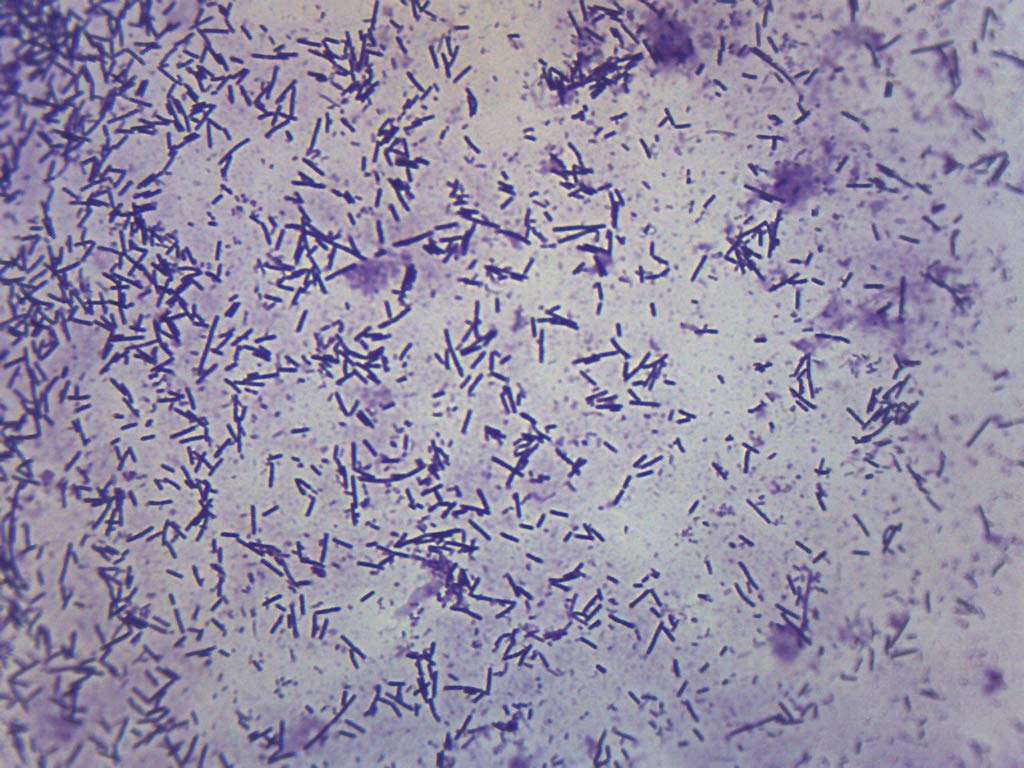


Escherichia Coli Smear Gram Stain Prepared Microscope Slide 75 X 25mm Biology Microscopy Eisco Labs Amazon Co Uk Business Industry Science
6 Now put under the microscope and observe at 100X by applying oil over slide Results & Observations Saureusà Methylene Blue Ecoli àSafranine BSubtilisàCrystal V àcoccus àrod àrod àbluepurple àpurple àvioloet 40X 40X 40X ScerevisiaeàTWM Pvulgaris àHDT àBrownian motion àflagellar motion àcolorless àcolorless 100X 100XThe prepared microscope slide image of Bacillus Subtilis at left was captured at 400x magnification Learn more here E Coli E Coli under the microscope at 400x E Coli (Escherichia Coli) is a gramnegative, rodshaped bacterium Most E Coli strains are harmless, but some serotypes can cause food poisoning in their hosts1 Before you plug in the microscope, turn the light intensity control dial on the righthand side of the microscope to 1 Now plug in the microscope and turn it on (see Fig 6) 2 Place a rounded drop of immersion oil on the area of the slide that is to be observed under the microscope, typically an area that shows some visible stain


What Does An E Coli Bacteria Look Like Under A Microscope Quora
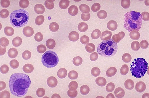


How These 26 Things Look Like Under The Microscope With Diagrams
Each group need to learn a simple brightfield microscope correctly by using at least three specimen slide with four objectives wit magnifications 0f 4x, 10x, 40x, 100x First, by using Escherichia coli (Ecoli), we start at 4x objective, followed by low power 10x objective viewing and the specimen looked clearer at 40x magnificationFirst, by using Escherichia coli (Ecoli), we start at 4x objective, followed by low power 10x objective viewing and the specimen looked clearer at 40x magnificationThe shaped of Ecoli is rodshaped and it is a gramnegative bacteriaThe gram stain of some specimen either gramnegative or grampositive can be resulted by looked at their color whether the bacteria are pink or red for gramMobile Game Studio Speacialized in Android, iOS , AR/VR Games Make your sketches as accurate as possible Make sure to give each figure a number and title (ex Figure 1 Onion Cell) magnification (40x, 100x, or 400x) label all visible cell parts *Use pencil or colored pencil ANIMAL CELLS RESULTS Microscope Observations *Within the circles below, draw what you see Find the perfect Plant


Microscopy Gram Staining Microscope World Blog


Microscope World Blog June 15
Entamoeba coli E coli cysts in concentrated wet mounts Cysts of Entamoeba coli are usually spherical but may be elongated and measure 10–35 µm Mature cysts typically have 8 nuclei but may have as many as 16 or more Entamoeba coli is the only Entamoeba species found in humans that has more than four nuclei in the cyst stage The nuclei may be seen in unstained as well as stainedE coli is one of the key prokaryotic model organisms used in the fields of biotechnology and microbiology Hence, in many recombinant DNA experiments, E coli serves as the host organism The reasons behind using E coli as the primary model organism are some characteristics of E coli such as fast growth, availability of cheap culture media to grow, easiness to manipulate, extensive5 Focus on the line with the 10X objective – refer to the microscope focusing procedure described in lab 1 Once you have focused on the specimen using the 10X objective, move the 40X objective lens into position Use the fine adjustment knob to bring the specimen into focus Now use the following
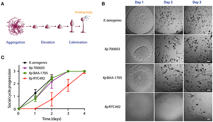


Frontiers Evaluating Different Virulence Traits Of Klebsiella Pneumoniae Using Dictyostelium Discoideum And Zebrafish Larvae As Host Models Cellular And Infection Microbiology


Tech Tip Imaging Bacteria Using Agarose Pads Biotium
B What organelles are present in E coli?Escherichia is a gramnegative bacterium, which under the microscope is shaped like a rod with a small tail It is widely distributed in nature (Brooker 08) Escherichia coli (E coli) is part of the normal intestinal flora Some strains are pathogenic and can cause gastroenteritis, UTI, meningitis, and wound infectionsSwitch to the MICROSCOPE tab to observe the sample as it would appear under the microscope By default, A rectangular piece of glass upon which a sample is mounted for viewing under a microscope 2 Manipulate With 40x selected, Click on the cow and observe E coli under the highest magnification
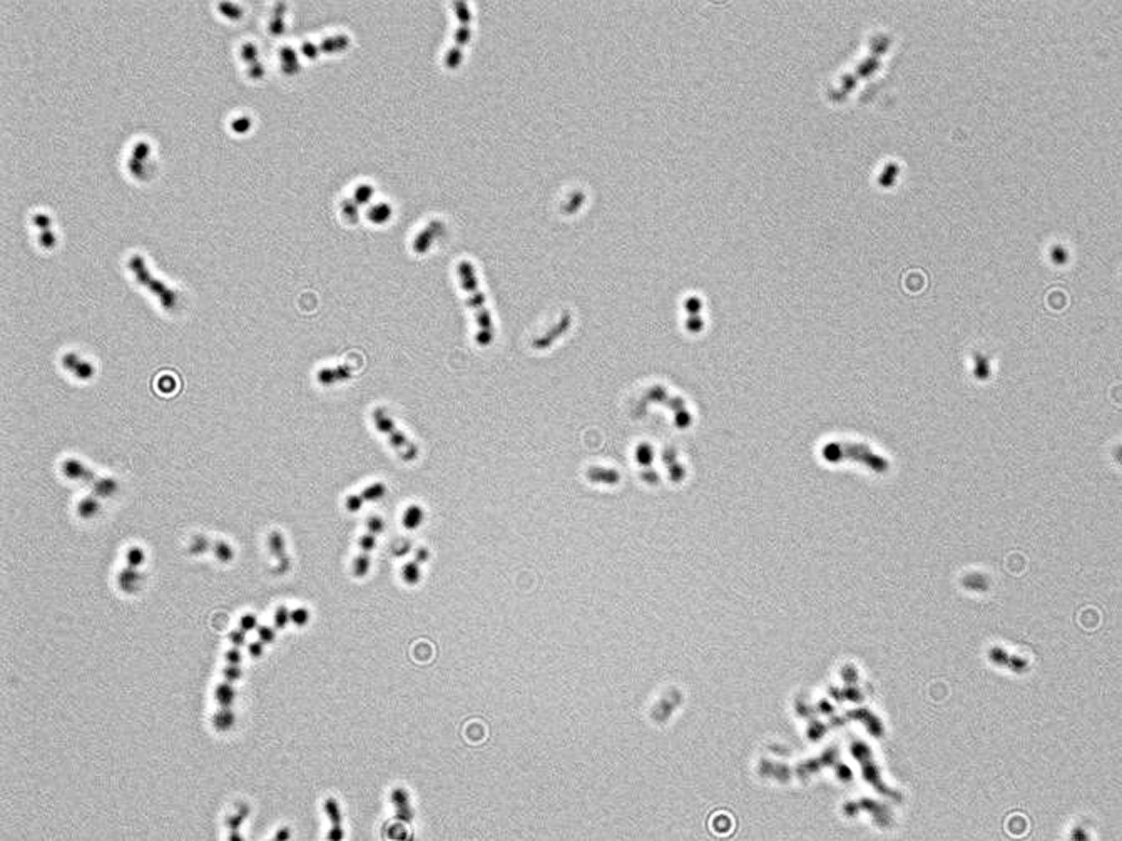


Microscopy For The Winery Viticulture And Enology


Morphology Of E Coli Cells Under Microscope At 100 Magnification Download Scientific Diagram
Step is repeated again with the 40X magnification although using the fine adjuster is accepted her as well Now before switching to the 100X magnification, apply a drop of immersion oil onto the part of the slide directly under the lens Now, using the fine adjuster ONLY, focus in onto your organism Those of you using E coliA rectangular piece of glass where a sample is mounted for viewing under the microscope What is the eye piece?7 NOW, switching to the 40X objective with the 10X eyepiece gives 400X magnification The field of view is This is illustrated by the 400X circle on the 100X view This circle is ¼ the diameter of the 100X view 8 Switch to the 400X view and 400X ruler (a millimeter ruler as seen under 400 power magnification)
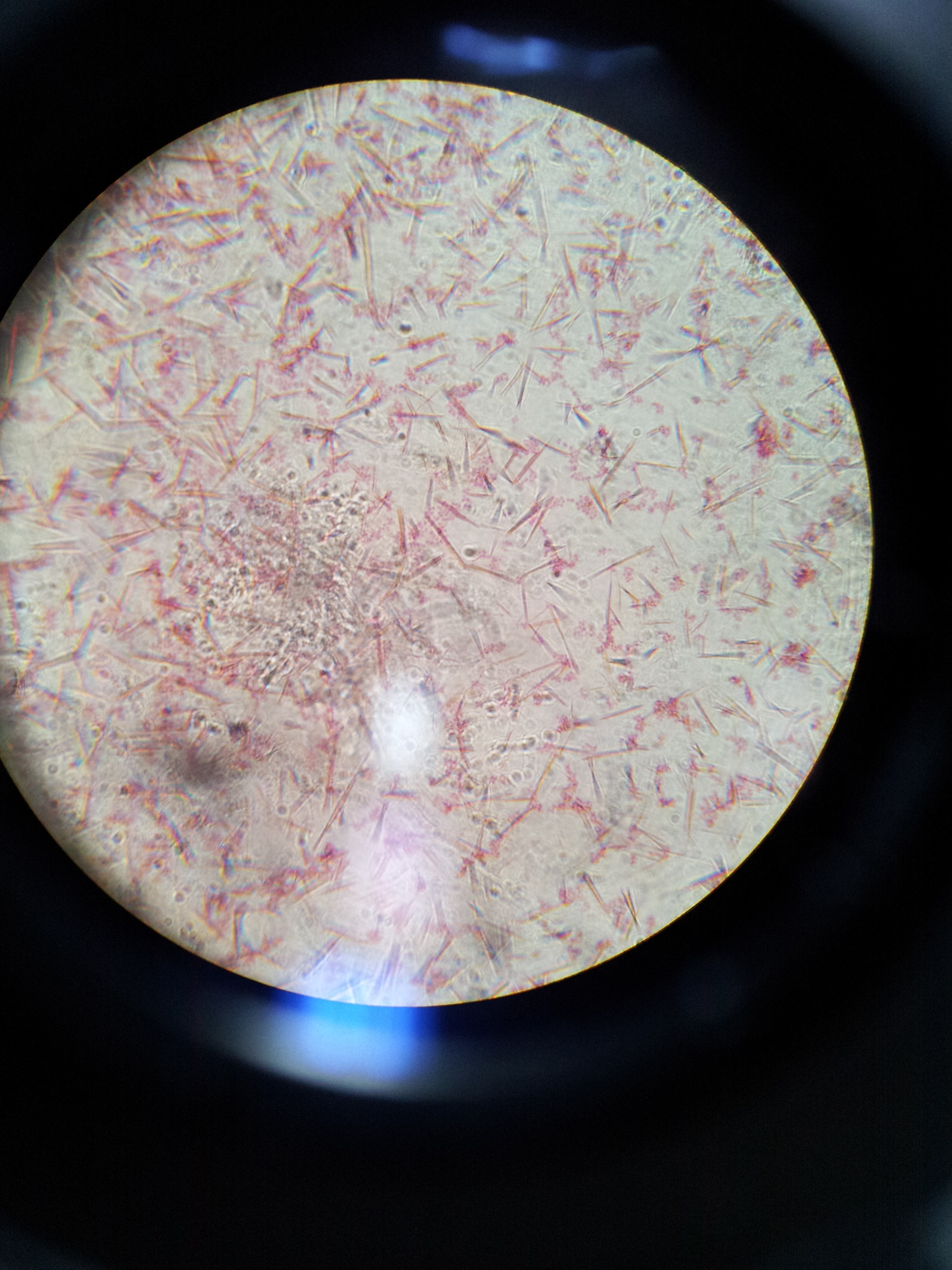


Lab 1 Principles And Use Of Microscope Ibg 102 Lab Reports


Microscopic Photograph Of The Microalgae Chlorella Vulgaris Used In The Download Scientific Diagram
4 Can a light microscope see bacteria?
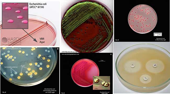


Escherichia Coli E Coli An Overview Microbe Notes


Microscopy And Staining
.jpg)


Escherichia Coli 400x Escherichia Coli 400x Manufacturers Escherichia Coli 400x Suppliers Escherichia Coli 400x Exporters Escherichia Coli 400x In India


A Control A Slide Showing Intact Rod Shaped E Coli 0157 H7 Cells And Download Scientific Diagram


Laboratory Test 1 Flashcards Chegg Com


Microscopic Studies Of Various Organisms


How These 26 Things Look Like Under The Microscope With Diagrams


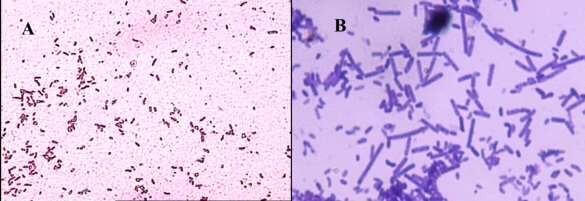
How Bacillus Spores Look Like Under Light Microscope


Observing Bacteria Under The Microscope Gram Stain Steps Rs Science


Cell Division Of E Coli With Continuous Media Flow Youtube


Q Tbn And9gcqkye60ou Johpr02n Mbv1fferrjpdh Lnct7ymdf5qhyia1ld Usqp Cau


Magnification Bioninja


Aph162 Report 1


Microscopic Studies Of Various Organisms


Zkfaa Bioproses Lab 1 Principles And Use Of Microscope


Entamoeba Coli Best View In Stool Microscopy At 40x E Coli In Stool Microscopy Parasites In Stool Youtube


Escherichia Coli E Coli An Overview Microbe Notes


Vade Mecum Microscope Practical Microscopy To Take With You Page 7


M60 Like Metalloprotease Domain Of The Escherichia Coli Yghj Protein Forms Amyloid Fibrils


What Does An E Coli Bacteria Look Like Under A Microscope Quora


Moving E Coli 1000 Magnification Youtube
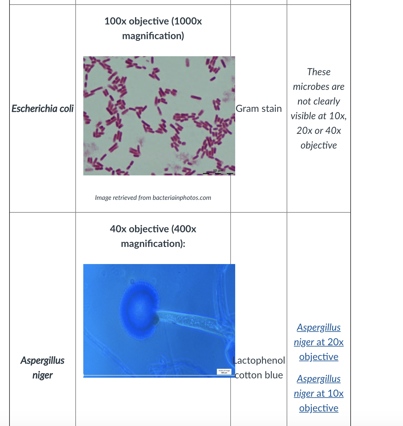


Solved Scientific Name Of Microorganism Pseudomonas Pseu Chegg Com


Lab 1


Staphylococcus Aureus And Ecoli Under Microscope Microscopy Of Gram Positive Cocci And Gram Negative Bacilli Morphology And Microscopic Appearance Of Staphylococcus Aureus And E Coli S Aureus Gram Stain And Colony Morphology On Agar Clinical


Gram Stain


Macroscopic And Microscopic Alterations Of Free Living Columbina Download Scientific Diagram


What Does An E Coli Bacteria Look Like Under A Microscope Quora
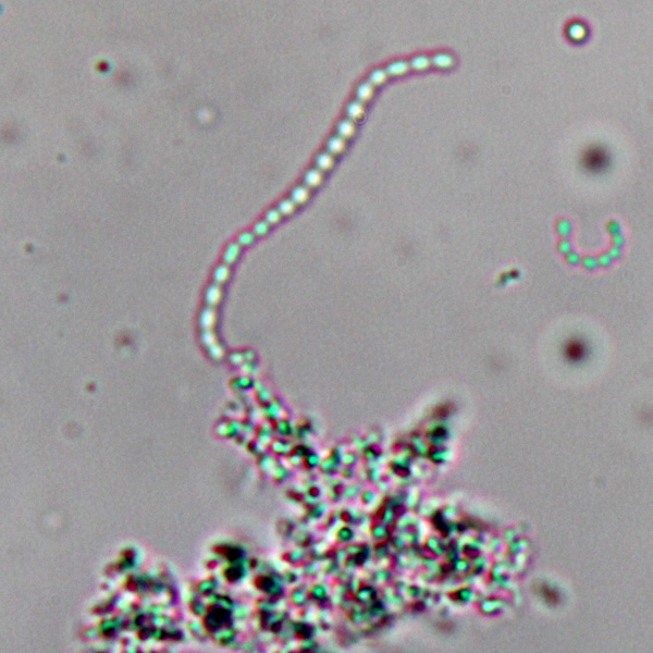


Observing Bacteria Under The Light Microscope Microbehunter Microscopy


Escherichia Coli Bacteria E Coli Stock Footage Video 100 Royalty Free Shutterstock


Escherichia Coli Bacteria E Coli Stock Footage Video 100 Royalty Free Shutterstock


Http Coltonanderson1 Weebly Com Uploads 2 4 3 0 Manual Pdf
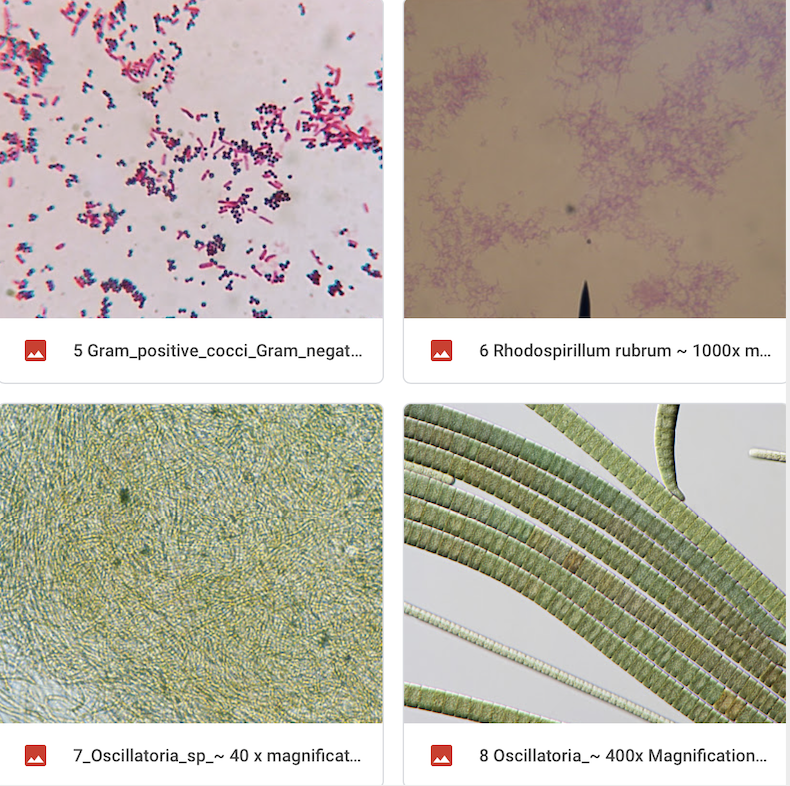


Solved Look At The 2 Gram Stain Slide This Is A Gram Sta Chegg Com
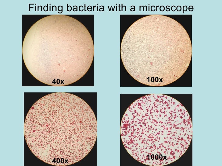


Chapter 3 Tools Of The Laboratory E Mail


Escherichia Coli E Coli An Overview Microbe Notes


Microscope World Blog Microscopy Gram Staining


Staphylococcus Aureus And Ecoli Under Microscope Microscopy Of Gram Positive Cocci And Gram Negative Bacilli Morphology And Microscopic Appearance Of Staphylococcus Aureus And E Coli S Aureus Gram Stain And Colony Morphology On Agar Clinical


Gut Bacteria Escherichia Coli Under Microscope Youtube


E Coli Gram Stain Page 1 Line 17qq Com


Escherichia Coli Light Microscopy


Www Entomoljournal Com Archives 17 Vol5issue6 Partac 5 4 293 708 Pdf


Free Living Amebae Media For Acanthamoeba And Naeglaria Cultures


Biol 230 Lab Manual Lab 1



Escherichia Coli Bacteria E Coli Stock Footage Video 100 Royalty Free Shutterstock


Aph162 Report 1


Bacillus Wikipedia


Gram Stain


E Coli Under The Microscope Types Techniques Gram Stain Hanging Drop Method


E Coli Gram Stain Page 6 Line 17qq Com


Q Tbn And9gcstf8rlsv6xf Tlmjmgapkp5vctdrjr4c7vz0kjmptie3nty0xo Usqp Cau


Observing Bacteria Under The Light Microscope Microbehunter Microscopy


E Coli Gram Stain Page 6 Line 17qq Com


E Coli Gram Stain Page 1 Line 17qq Com


Bright Field Microscopy An Overview Sciencedirect Topics


Biotek Instruments Image Of The Week E Coli Expressing Gfp At 40x Imaging Microscopy


Gram Stain Staphylococcus Aureus And Escherichia Coli Gram Staining Technique Micrograph Of S Aureus And E Coli


E Coli Under The Microscope Types Techniques Gram Stain Hanging Drop Method


Bacteria Under The Microscope E Coli And S Aureus Youtube


Does Spiral Bacteria Produce Endoospore


Gram Stain


Image Result For Negative Stain E Coli Bacillus Microbiology Stain
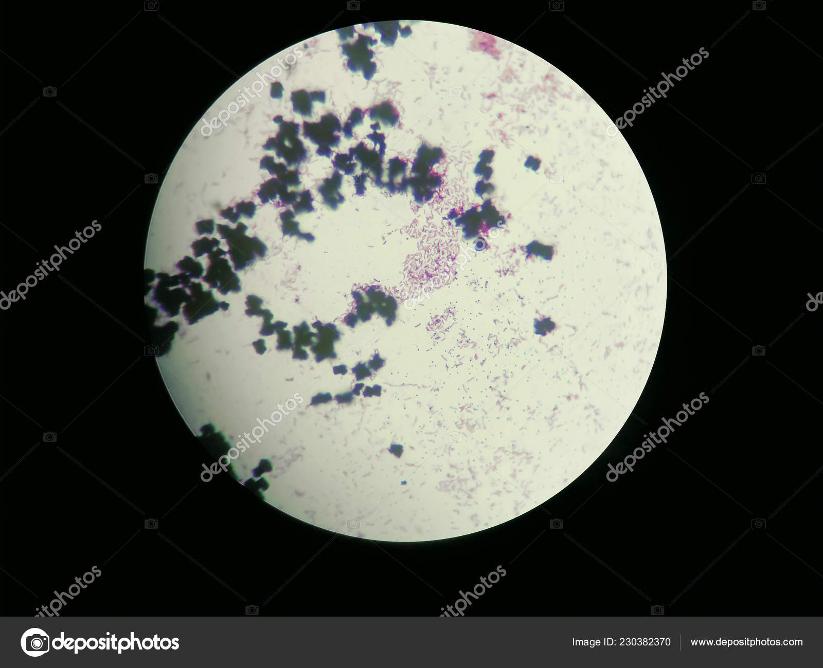


Image Of Escherichia Coli Obtained Through A Light Microscope Stock Photo Image By C Pike 28


Gram Stain
.jpg)


Escherichia Coli 400x Escherichia Coli 400x Manufacturers Escherichia Coli 400x Suppliers Escherichia Coli 400x Exporters Escherichia Coli 400x In India


Plos One Treatment With High Dose Antidepressants Severely Exacerbates The Pathological Outcome Of Experimental Escherichia Coli Infections In Poultry


Aph162 Report 1


Biol 230 Lab Manual Lab 1


What Does An E Coli Bacteria Look Like Under A Microscope Quora
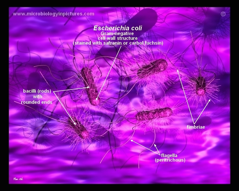


How E Coli Bacteria Look Like


Dynamic Persistence Of Intracellular Bacterial Communities Of Uropathogenic Escherichia Coli In A Human Bladder Chip Model Of Urinary Tract Infections Biorxiv


Lab Manual Exercise 1


Www Mccc Edu Hilkerd Documents Bio1lab3 Exp 4 Pdf


Biol 230 Lab Manual Lab 1


コメント
コメントを投稿